Identifying asbestos: can you tell from a photo?
We had five samples from Burgenland analysed in a laboratory. Guess for yourself first: which rock, which asbestos minerals, what percentage. Then we show what the lab found. The answer up front: you cannot tell from a photo alone. That is exactly the point.
Your guesses, compared with everyone playing
You have guessed all five samples. The overview below shows your guesses next to the aggregated guesses of everyone who has played so far. The lab answer is on each sample's card directly.
| Sample | Rock, you | Rock, crowd majority | Minerals, you | Minerals, crowd top | Asbestos content, you | Asbestos content, crowd mean |
|---|
Crowd values are updated after each time you save. Majorities and means are based on the n of guesses so far; it is shown in brackets in the first column for each sample.
The answer in detail
What the lab saw
From the photo it could not be told; under the scanning electron microscope it could. Here are the actual fibres of each sample, with the verdict. Click an image to zoom in at full resolution.
Sample 001over 50%
Tremolite, over 50%. Under the scanning electron microscope, a dense felt of straight, stiff amphibole fibres.
Sample 002over 50%
Tremolite, over 50%. Splaying fibre bundles, typical of amphibole asbestos.
Sample 003traces
Chrysotile, only traces. The only asbestos form of the serpentine group, here a single curved, flexible fibre in the granular material: the needle in the haystack.
Sample 004over 50%
Tremolite, over 50%. A dense, parallel-aligned bundle of straight, prismatic needles.
Sample 0061 to 5%
Anthophyllite and tremolite, 1 to 5% each. A mixed finding of two amphiboles, straight rod-shaped fibres between mineral grains.
What is asbestos, and why Burgenland in particular?
Asbestos is a collective term for six fibrous-crystallising minerals: chrysotile (serpentine group) and actinolite, tremolite, anthophyllite, amosite, and crocidolite (all amphiboles). When inhaled, the fine, barely degradable fibres lodge permanently in lung tissue. After a long latency of often 20 to 40 years, they can give rise to pleural plaques, lung cancer, and the usually fatal mesothelioma. There is no safe threshold; the risk rises linearly with the cumulative fibre dose.
Most people associate asbestos with industrial products: fibre-cement (Eternit) sheets, old floor adhesives, gaskets. In Burgenland, however, there is additionally a natural occurrence. In the so-called Rechnitz Window, a geological structure on the Austrian-Hungarian border, a strip of metamorphic rock comes to the surface beneath the nappes of the Eastern Alps. Among these rocks is serpentinite: a dark-green rock formed from the mantle minerals olivine and pyroxene, which can hold chrysotile veins in its fractures. Immediately adjacent are amphibole-bearing rocks whose fibrous forms are actinolite or tremolite asbestos.
This very rock was quarried at sites such as Pilgersdorf, Bernstein, Rumpersdorf, and Badersdorf, crushed, and put into circulation as grit, chippings, gravel, and milled material. What cannot be read from this material is its asbestos content. If you have a photo of a chunk, you see a piece of rock. If you have the laboratory microscope, you see the fibres. The difference cannot be guessed, only measured.
Mineralogy
Two families, two fibre shapes
Asbestos is not a single mineral but a collective term for two mineralogical families with different crystal structures and visibly different fibre shapes: the serpentine group with chrysotile, and the amphiboles, in Burgenland above all tremolite, actinolite, and anthophyllite.
Serpentine: chrysotile sheet silicate · Mg₃Si₂O₅(OH)₄
flexible
wavy
Mg silicate
Chrysotile is the only asbestos mineral of the serpentine group. Its sheets are curved and roll up into fine tubes. Exactly this curvature turns the sheet silicate into a flexible, wavy fibre, the asbestiform form of serpentine, known industrially as "white asbestos".
Source: Evans (2004), summarised in our geology article.Amphiboles: tremolite, actinolite, anthophyllite a different mineral family · straight needles
straight
stiff
prismatic
The amphibole asbestos minerals, in Burgenland above all tremolite, actinolite, and anthophyllite, form a different mineral family. They grow not wavy but as straight, stiff, prismatic needles. Under the scanning electron microscope they appear as straight needles that splay into finer fibrils at their ends, clearly different from the wavy chrysotile fibres.
Sources: Van Gosen and Clinkenbeard (2011); Koller (1985), summarised in the geology article; SEM images: CRB Analyse Service GmbH (test report 26-07931).When fine enough, fibres of both families meet the WHO fibre geometry (length over 5 µm, diameter under 3 µm, ratio over 3:1) and are then considered respirable. Amphibole asbestos is epidemiologically considered more potent than chrysotile; we place the evidence and its limits in context on the standards page (section 5). How asbestos forms in serpentinite at all is covered in detail in the geology article.
Marked in the image
Same word, two shapes
The same SEM images as above, this time labelled: the straight, stiff needles of an amphibole (tremolite, sample 004) alongside the single curved fibre of serpentine (chrysotile, sample 003). Switch the labels on.
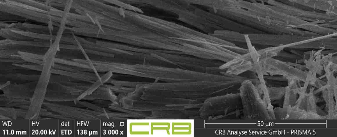
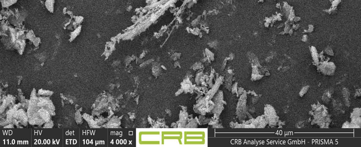
Both are asbestos, but the shape separates the families: amphiboles grow as straight, stiff needles, chrysotile as a curved, flexible fibre. Fine enough, both meet the WHO fibre geometry (length over 5 µm, diameter under 3 µm, ratio over 3:1) and count as respirable. More on this in the geology article and on the standards page.
SEM images: CRB Analyse Service GmbH, test report 26-07931.Size comparison
How small is a WHO fibre?
The dangerous fibres are invisible to the naked eye. To scale, from top to bottom: a human hair, the fine dust PM10, and the diameter of a WHO fibre. Tap a bar.
Across the width of an average hair (about 70 µm), more than 20 WHO-fibre diameters fit side by side.
Method: EDX
The elemental fingerprint
Under the scanning electron microscope, the shape reveals that a fibre is present. Which mineral it is, the elemental analysis (EDX) shows: each mineral leaves a characteristic set of X-ray lines. That is how the lab tells the asbestos families apart. Choose a spectrum and tap the peaks.
Tip: hover over a peak or tap it to see the element.
Reading aid: calcium (3.7 keV) marks tremolite, iron (6.4 keV) anthophyllite; chrysotile is magnesium-rich and calcium-free. The gold peak (Au) comes from the thin sputter coating that makes the sample conductive in the SEM, and is not part of the mineral. How the lab counts and classifies fibres is on the standards page.
Source: CRB Analyse Service GmbH, test report 26-07931, six measurement points (spectra ES1 to ES6). The charts are redrawn from the measured peak heights; the heights are relative within each spectrum.Mini-game
Can you identify the mineral from its fingerprint?
You have seen the three signatures. Now the other way around: here is a spectrum, which mineral is behind it? Watch for calcium (tremolite), for iron (anthophyllite), or for a magnesium-rich, calcium-free spectrum (chrysotile).
Sample preparation
From chunk to image
How does a piece of road gravel become a finding? The sample is crushed, applied as a thin dispersed preparation on a holder, imaged in the scanning electron microscope, and analysed by EDX. These four photos show the preparation in the lab.
-
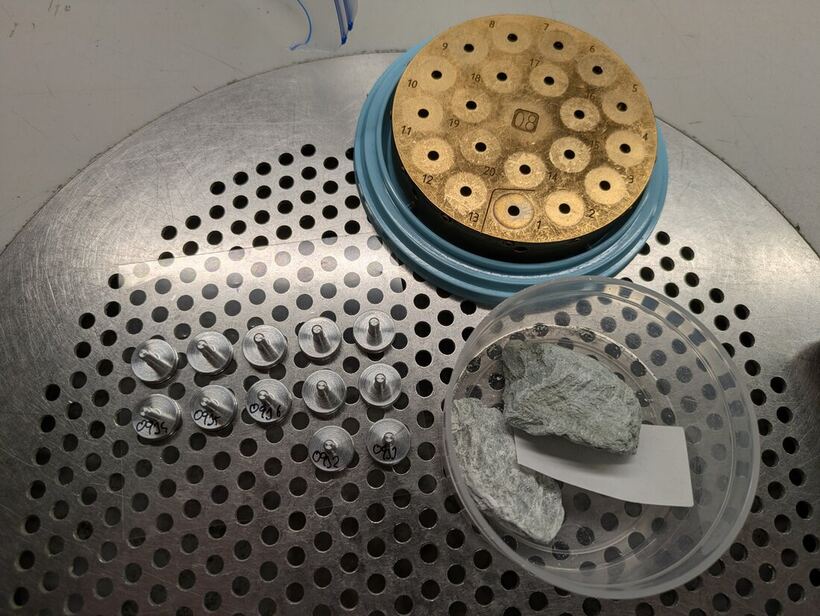
A Burgenland sample, a grey-green rock chunk, next to the stage with the carbon sample holders. -
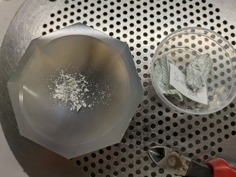
The loose sample material next to the chunk, ready for crushing. -
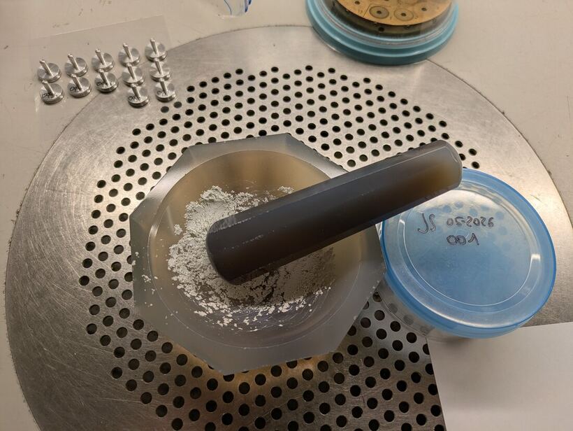
Crushing in an agate mortar to a fine powder. -
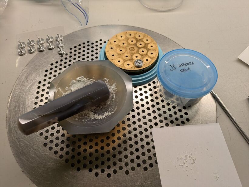
Applying it as a dispersed preparation onto the sample holders. Imaging in the scanning electron microscope follows.
Videos
Asbestos in the field
Mag. Julia Stipsits documents the asbestos finds in Burgenland on site. Here is one of her videos; further footage from forest and trail will follow, as will short SEM zoom clips into the fibres.
SEM zoom clips to follow
Image credits and thanks
The sample photos on this page were provided by Mag. Julia Stipsits, who does essential awareness-raising work on the asbestos problem in Burgenland through her social-media channels. We thank her for providing the images and for her efforts on the ground.
Instagram: @MissJulsi · TikTok: @MissJulsi
As of 18 June 2026. This page was revised: the lab results (CRB Analyse Service GmbH, test report 26-07931) are published, together with SEM and EDX images, a size comparison, and interactive explainers. The guessing game now shows the five samples measured in the lab.
📄 Download test report 26-07931 (PDF, 8 pages). CRB Analyse Service GmbH, DAkkS-accredited, method VDI 3866 Part 5.